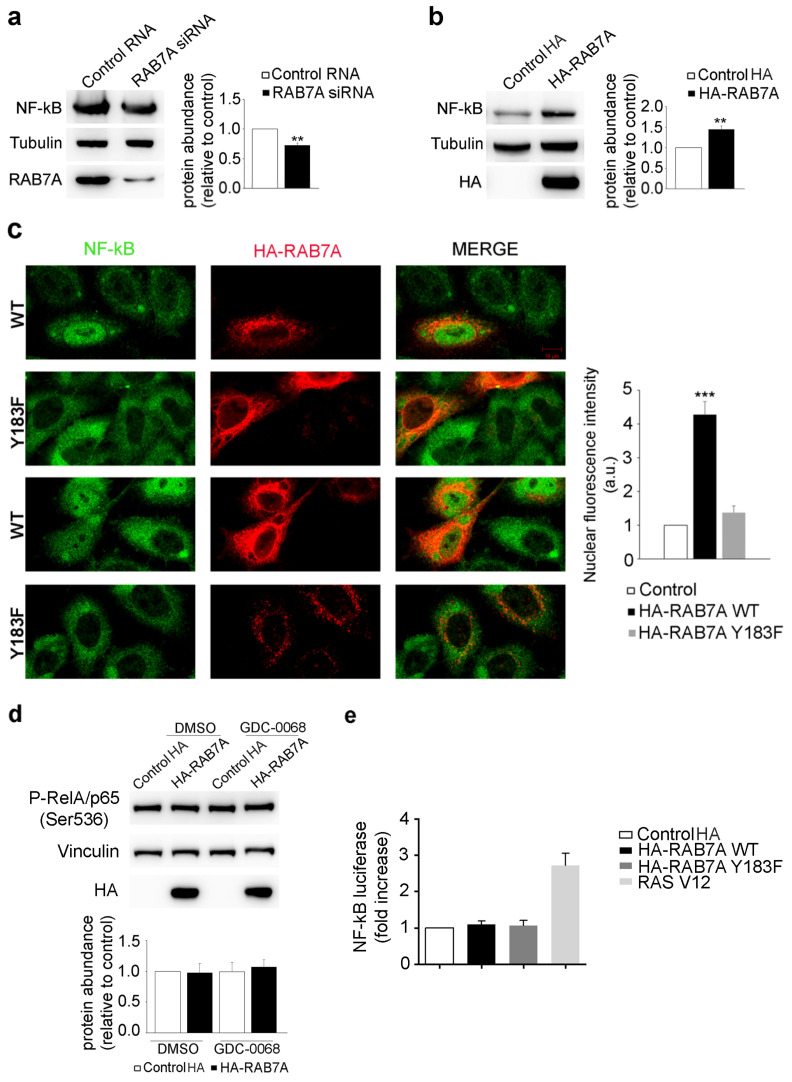
Figure 6.
RAB7A affects NF-kB expression and localization. (a) HeLa cells were transfected with either control RNA or RAB7A siRNA, replated 72 h after transfection and then lysed after 48 h. (b) HeLa cells were transfected with plasmids encoding either HA or HA-RAB7A and lysed after 24 h. Lysates were subjected to Western blot analysis using anti-NF-kB, anti-tubulin, anti-RAB7A and anti-HA antibodies. The quantification of NF-kB is shown. Data represent the mean ± s.e.m. of at least three independent experiments. (c) HeLa cells were transfected with a plasmid coding for HA-RAB7AWT or HA-RAB7AY183F and, 24 h after transfection, were analyzed by immunofluorescence using anti-HA (red) and anti-NF-kB (green) antibodies. Scale bar = 10 µM. NF-kB nuclear fluorescence intensities of at least 50 cells per sample were measured using ImageJ. Data represent the mean ± s.e.m. of three independent experiments. Statistical analyses were performed using Student’s t test with control cells as referring sample. (d) HeLa cells were transfected with plasmids encoding either HA, HA-RAB7AWT or HA-RAB7AY183F. Then, 24 h after transfection cells were treated with AKT inhibitor GDC-0068 1 µM or DMSO for 24 h and then lysed. Lysates were subjected to Western blot analysis using anti-P-p65/RelA (Ser536), anti-vinculin and anti-HA antibodies. (e) HeLa cells were transfected with the NF-kB luciferase reporter vector and with different expression vectors. Twenty-four hours after transfection, cells were lysed and the luciferase activity was measured in cell extracts. Data are represented as fold induction of the normalized luciferase activity with respect to control cells transfected with GFP. All luciferase results represent the average ± S.D. of three independent experiments. All samples were read in triplicate. ** = p < 0.01; *** = p < 0.001.

