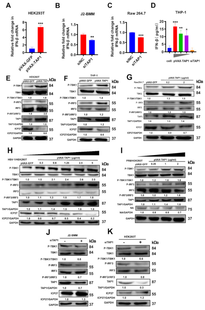
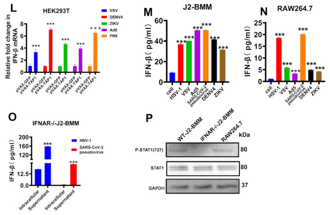
Figure 4.

TAP1 significantly promoted the interferon (IFN)-β production through activating the TANK binding kinase-1 (TBK1) and the interferon regulatory factor 3 (IRF3) signaling transduction. (A) HEK293T cells were transfected with plasmids expressing GFP or TAP1 (1 μg/mL) for 24 h, respectively, followed by HSV-1 infection (MOI = 0.25) for 8 h. Then, the expression of IFN-β was quantified by RT-qPCR. (B) WT-J2-BMM cells were transfected with siNC, siTAP1 for 24 h, followed by HSV-1 infection (MOI = 0.25) for 8 h. Then, the expression of IFN-β was quantified by RT-qPCR. (C) Raw264.7 cells were transfected with siNC, siTAP1 for 24 h, followed by HSV-1 infection (MOI = 0.25) for 8 h. Then, the expression of IFN-β was quantified by RT-qPCR. (D) THP-1 cells were transfected with TAP1-expressing plasmid in different concentration (5 μg/mL, 1.25 μg/mL, 0.3 μg/mL) for 24 h, followed by HSV-1 infection (MOI = 0.25). Then, the expression of IFN-β in cell lysates was determined by ELISA kits. (E–G) HEK293T, THP-1 or Raw 264.7 cells were transfected with plasmids expressing GFP or TAP1 (1 μg/mL) for 24 h respectively, followed by HSV-1 infection (MOI = 0.25) for 8 h. Then, the expression of targeted proteins was detected by Western blot. The relative ratios of targeted protein and GAPDH were marked at the bottom of the pictures. (H,I) HEK293T cells were transfected with plasmids expressing GFP or TAP1 for 24 h, followed by viral infections (HSV-1 or PR8) for 24 h at MOI of 1. Then, the expression of targeted proteins was detected by Western blot. The relative ratios of targeted protein and GAPDH were marked at the bottom of the pictures. (J,K) WT-J2-BMM or HEK293T cells were transfected with siNC or siTAP1 for 24 h, followed by HSV-1 infection (MOI = 0.25) for 8 h. Then, the expression of targeted proteins was detected by Western blot. The relative ratios of targeted protein and GAPDH were marked at the bottom of the pictures. (L) HEK293T cells were transfected with or without TAP1-expressing plasmid for 24 h, followed by viral infections for 8 h at MOI of 1, including VSV, ZIKV, DENV4, Ad5 and PR8. Then, the expression of IFN-β was quantified by RT-qPCR. (M,N) WT-J2-BMM or Raw 264.7 cells were transfected with TAP1-expressing plasmid for 24 h, followed by viral infections for 24 h at 1 MOI, including HSV-1, VSV, Ad5, SARS-CoV-2 pseudovirus, ZIKV, DENV4. Then, the expression of IFN-β in cell lysates was determined by ELISA kits. (O) (IFNAR−/−)-J2-BMM cells were transfected with TAP1-expressing plasmid for 24 h, followed by HSV-1 or SARS-CoV-2 pseudovirus infection for 24 h. Then, the expression of IFN-β in cell lysates and supernatant was determined by ELISA kits. (P) WT-J2-BMM, (IFNAR−/−)-J2-BMM, and Raw 264.7 cells were transfected with TAP1-expressing plasmid for 24 h, followed by HSV-1 infection for 24 h. Then, the level of STAT1 phosphorylation and total STAT1 was measured by Western blot. The data from at least triplicates were shown as the mean ± SD. * p < 0.05, ** p < 0.01 and *** p < 0.001.

