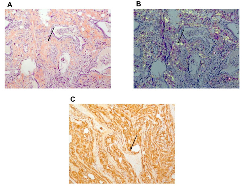
Figure 6.
Histology: thyroid pathologic sample of patient B with amyloid deposits. (A) Congo red staining showing amyloid deposits. (B) Typical yellow-green bi-refringence of amyloid deposits under polarized light showing amyloid deposits. (C) Immuno-histochemistry. Brown staining due to strong fixation of anti-SAA antibody by amyloid deposits. Black narrow indicates amyloid deposit.

