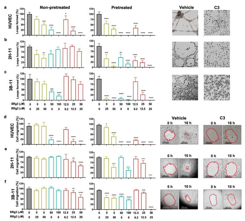
Figure 2.
Effect of Rg3 epimers on loop formation (a–c) and migration (d–f) of human umbilical vein endothelial cell (HUVEC), 2H-11, and 3B-11 cells. Analysis of the loop formation and migration was performed at two timepoints; non-pretreated cells and 3-day pre-treated cells. Treatments are shown on the x axis, and (a–c) show the results of the loop formation in the HUVEC, 2H-11, and 3B-11 cell lines, respectively, at peak loop formation timepoints—16 h for HUVEC and 4 h for 2H-11 and 3B-11 cells. The experiments were done in triplicate and the results are presented as mean ± standard deviation (SD; p < 0.05). (d–f) show the results of cell migration in HUVEC, 2H-11 and 3B-11 cell lines, respectively. Results are presented as mean ± SD of 3 and 6 replicates for loop formation and migration assays, respectively (p < 0.05). The images represent the pre-treated cells. C3 represents a combination of 50 µM SRg3 + 25 µM RRg3. * p < 0.05, ** p < 0.01, *** p < 0.001 and **** p < 0.0001.

