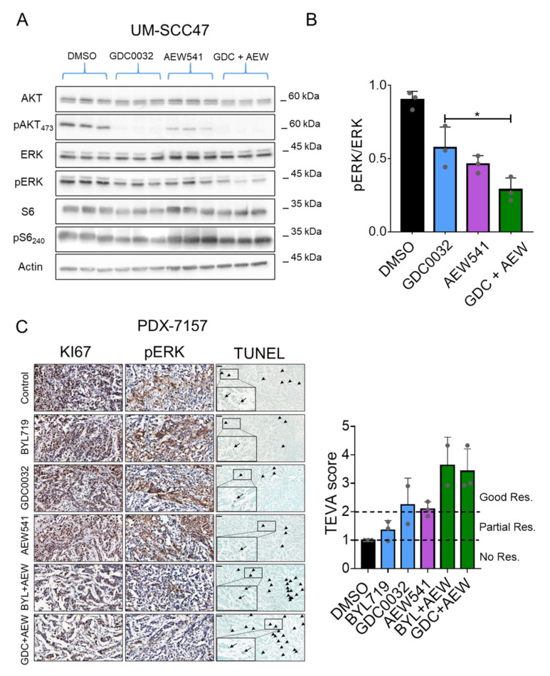
Figure 5.
isiPI3K and AEW541 induced growth in a PDXHPV+ ex vivo model. (A) UM-SCC47 sensitive cells were treated for 4 h with DMSO, GDC0032 (500 nM), AEW541 (1 μM), or a combination of the two drugs, after which cells were lysed. The indicated proteins and phosphorylated proteins were measured using western blot. (B) pERK plot was quantified with Adobe Photoshop 2020and normalized to ERK. The western blotting experiment was repeated 3 times, and representative results from one experiment are presented. (C) PDXHPV+ was cultured with DMSO, GDC0032 (500 nM) or BYL719 (2 μM), AEW541 (1 μM), or a combination of AEW541 with GDC0032 or BYL719, as indicated, for 24 h, after which the tissue was fixed and analyzed using IHC. Representative images of IHC staining for KI67, pERK, and TUNEL are presented together with the TEVA score. Scale bar for KI67 and MAPK: 20 μm, for TUNEL: 50 μm. Statistical significance was calculated by one-way ANOVA, * p < 0.05.

