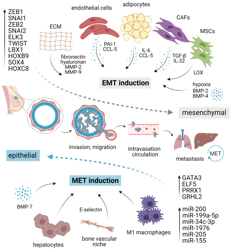
Figure 1.
EMT/MET plasticity in breast cancer. EMT is induced by pro-inflammatory cytokines and molecules secreted by different stromal cells present in the tumor microenvironment, ECM elements, and hypoxia. Different cell types such as macrophages or hepatocytes might activate MET. Each program is executed via a specific set of transcription factors and/or corresponding miRNAs. The activation of EMT and mesenchymal phenotype (gray) grants cancer cells the ability to migrate, invade, intravasate, survive in circulation, and extravasate at distant sites. At the secondary organs, mesenchymal cells revert to an epithelial state (blue) through MET, regaining the ability to form macrometastasis. EMT, epithelial–mesenchymal transition; MET, mesenchymal–epithelial transition; CAFs, cancer-associated fibroblasts; MSCs, mesenchymal stromal cells; ECM, extracellular matrix; MMP, matrix metalloproteinase; LOX, lysyl-oxidase. Created with BioRender.

