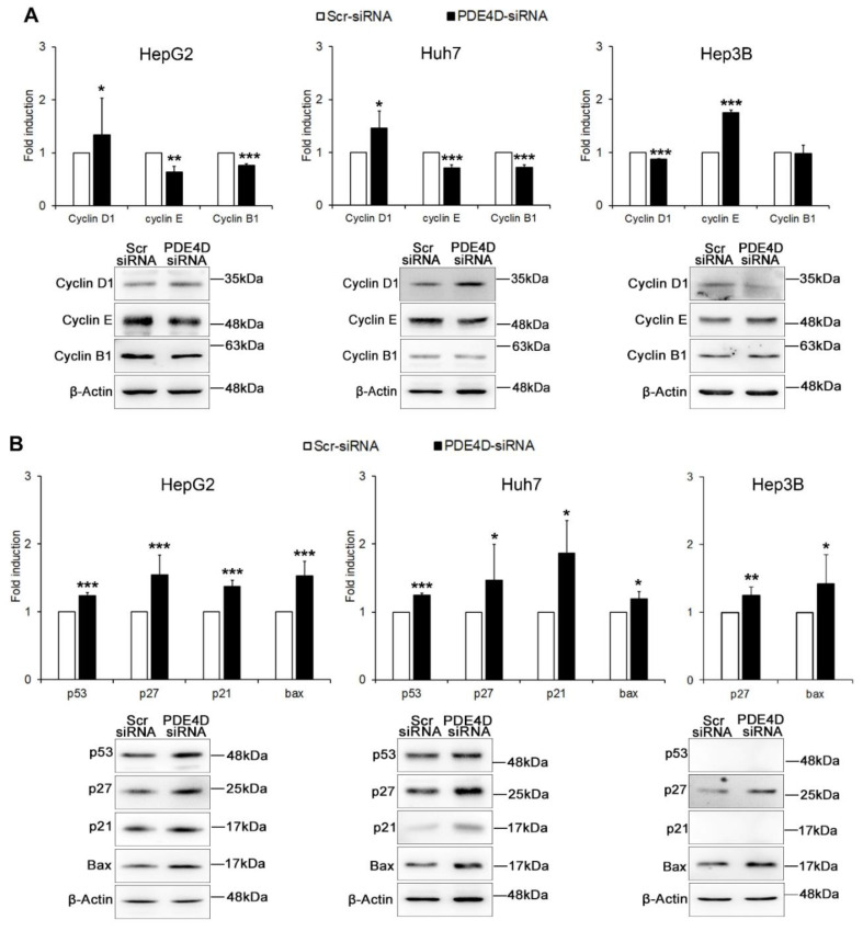
Figure 4.
The effect of PDE4D gene silencing on cell cycle regulators and apoptotic proteins. Western blot analysis in HepG2, Huh7 and Hep3B cells 48 h after transient silencing. The graphs represent densitometric quantifications of cyclins D1, E, and B1 (A), and of apoptosis regulators p53, 27, p21, Bax (B), all of proteins normalized to β-actin. Data are the mean ± SD of three independent experiments. Student t test. * p < 0.05; ** p < 0.01; *** p < 0.001 versus Scr-siRNA.

