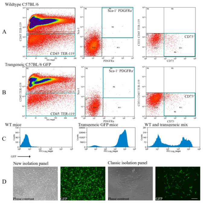
Figure 1.

Comparison between the classic FACS staining panel and the new protocol to isolate freshly purified MSCs. (A) Displays whole bone marrow cells from wild-type C57BL/6 mice. (B) Shows whole bone marrow cells from transgenic GFP C57BL/6 mice. Green squares indicate the isolated populations. The staining panel were either CD45 TER-119 Sca-1+ PDGFR-α+ (middle) or CD31− CD45 TER-119 CD73+ (right). (C) Exhibits the GFP signal in the wild-type (left), GFP transgenic (middle), and wild-type-transgenic mix (right, only for illustration). (D) Depicts isolated cells after 2 to 4 days of culture. The culture was only for illustrative purpose, not for transplantation. Scale bar = 100 µm, 200× magnification.
