
Figure 2.
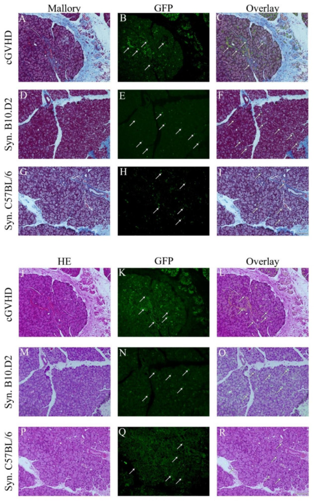
Mesenchymal stem cells become different phenotype after syngeneic and allogeneic MSC transplantation. Mallory (top A–I) and HE (bottom J–R) staining of the lacrimal gland in the allogeneic (cGVHD n = 19) and two syngeneic models (B10.D2 (n = 3) and C57BL/6 (n = 25) recipient mice). Scale bar = 100 µm, 200× magnification. Arrows indicate GFP+ cells. Mallory and HE staining are on the left side. The middle depicts the GFP channel and the right is a merge of both.