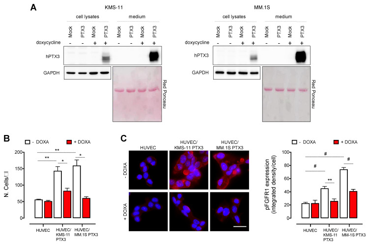
Figure 1.
PTX3 released by MM cells impairs HUVEC proliferation by inhibiting FGFR activation. (A) Western blot analysis of PTX3 expression (cell lysates) and release (medium) from KMS-11 and MM.1S cells transduced with a doxycycline (DOXA)-inducible PTX3 (PTX3) or a control vector (Mock) and treated or not with DOXA for 48 h. (B) Cell count by cytofluorimetric analysis of HUVEC co-cultured or not with KMS-11 PTX3 or MM.1S PTX3 cells for 48 h in the presence or absence of DOXA. (C) Left panel: Immunofluorescence analysis of phospho-FGFR1 (red fluorescence) expression in HUVEC co-cultured or not with KMS-11 PTX3 or MM.1S PTX3 cells for 24 h in the presence or absence of DOXA. Scale bar: 50 μm. Right panel: Fluorescence intensity quantification of phospho-FGFR1 by ImageJ software. For each microscopic field, fluorescence intensity values were normalized with the number of nuclei detected by DAPI staining. Data are mean ± SEM of 3 experimental replicates. * p < 0.05, ** p < 0.01, # p < 0.001.

