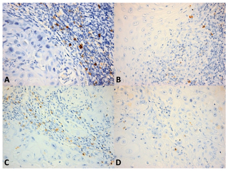
Figure 3.
CD56+ cells in OSCC. (A) Numerous CD56+ cells within the inflammatory infiltrate bordering the tumor invasion front; (B) Few CD56+ cells (less than eight cells/high power field) within the invasion front; (C) Numerous CD56+ cells within the intratumor inflammatory infiltrate; (D) Scattered isolated CD56+ cells within the tumor. CD56 × 400.

