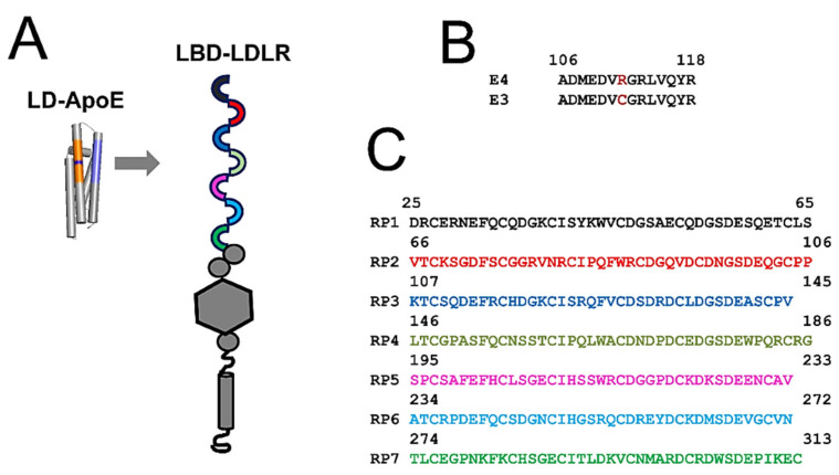
Figure 1.
Ligand domain (LD) of ApoE and LDL-A repeats of LDLR. (A) The secondary structure of ApoE-LD and the domain structure of LDLR are shown. The receptor-binding interface is a part of the fourth α-helix in the ApoE-LD domain (shown in blue). The R112C domain portion of the third α-helix in the ApoE-LD domain is shown in orange. Position 112 within the R112C domain is shown in purple. Seven amino-terminal LDL-A repeats are shown as colored semicircles. (B) Primary sequence alignment of R112C domains from ApoE4 and ApoE3 isoforms. Amino acid in position 112 is shown in red (R for ApoE4 and C for ApoE3). (C) Primary sequences of seven LDL-A repeats from the LDLR. The sequences of RP1–RP7 repeats are color-coded according to the panel A diagram.

