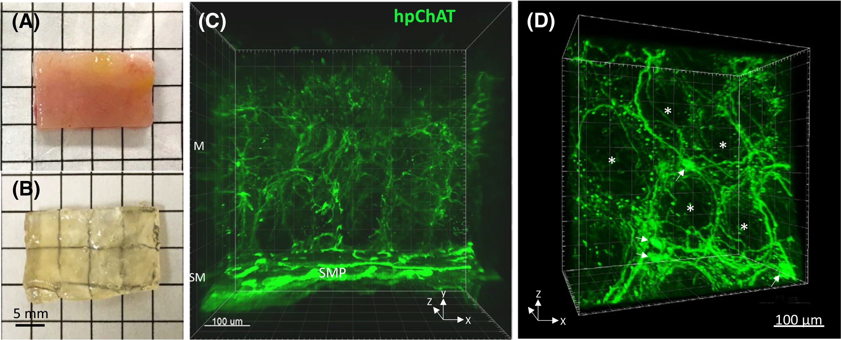
FIGURE 2.

A non-diseased sigmoid colon tissue before (A) and after (B) clearing with the modified passive CLARITY technique. The tissue-hydrogel hybrid turned transparent (B) and permeable for antibody penetration (C,D). C, A 3D image (500 μm depth scan) of the mucosa (M) and submucosa (SM) in the transparent tissue stained by immunofluorescence using a novel mouse antiserum against human peripheral choline acetyltransferase (hpChAT). Green fluorescent hpChAT-ir nerve fibers in the mucosa appeared to be projected from hpChAT-ir neurons in the submucosal plexus (SMP). A 360-degree panoramic presentation of this 3D image is shown in Video S1. D, A 3D Image in the lamina propria showing fine hpChAT-ir nerve fiber strands forming a honeycomb-like network around the mucosal crypts (*). A few nerve cell bodies (arrows) were seen at the intersections of nerve fiber strands
