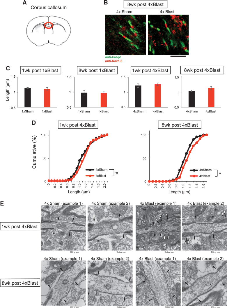
FIG. 10.
bTBI altered the structure of the nodes of Ranvier in the corpus callosum. (A) Illustration of a coronal section (bregma +0.5 mm) containing the corpus callosum region examined. (B) Representative images of nodes of Ranvier staining with anti-Caspr and anti-Nav1.6 antibodies at 8 weeks after 4 × Blast. Scale bar, 5 μm. (C) Quantification of nodal gap length for 1 × Blast (n = 7) and 4 × Blast (n = 7) at 1 and 8 weeks. n = 9 for 4 × Sham and n = 8 for 4 × Blast at 1 week, n = 7 for 4 × Sham and n = 6 for 4 × Blast at 8 weeks. (D) Cumulative percentage analysis of nodal gap length for 4 × Blast at 1 and 8 weeks. *p < 0.05, Kolmogorov-Smirnov test. n = 83, 107, 71, and 87 for 1 week after 4 × Sham, 4 × Blast, 8 weeks after 4 × Sham, 4 × Blast, respectively. (E) Electron micrographs of nodes of Ranvier in the corpus callosum for 1 week (top) and 8 weeks (bottom) after 4 × Sham and 4 × Blast mice. Filled arrowheads indicate the nodal gap and paranodal borders. Open arrowheads show distorted paranodal morphology in 4 × Blast mice. Scale bars, 100 nm for 1 week after 4 × Sham (example 2) and 8 weeks after 4 × Blast (example 2); 500 nm for all other images (embedded in original micrographs). bTBI, blast traumatic brain injury; Caspr, contactin-associated protein. Color image is available online.

