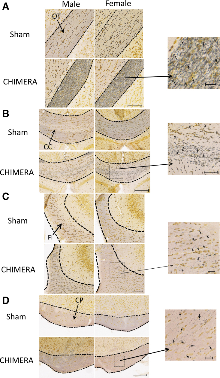
FIG. 2.
Axonal degeneration as observed by silver staining in the optic tracts (OT; A), corpus callosum (CC; B), fimbria (FI; C), and cerebral peduncles (CP; D). Scale bars in (A) and (C) represent 100 μm, (B) and (D) represent 200 μm. Photomicrographs on the right are enlarged images of the boxed regions; scale bars of enlarged regions represent 20 μm (A), (C) and (D), or 50 μm (B). Arrows in enlarged regions point to silver-stained punctate or argyrophilic fibers. All injured mice showed prominent silver staining in the OTs (A), and readily observable silver staining in the CC (B, with enlarged region). About one-third of mice (both male and female) had sparse silver staining in the FI, lateral to CA2, observable with higher magnification (C, enlarged region). Most injured animals also showed white matter damage in the CPs, which was only readily observable at higher magnification (D, enlarged region).

