
Table 2.
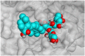
3D pictures representing both the binding modes and the positioning inside the protein pocket of the SARS-CoV-2 main protease between the best-selected triterpenes (SAP5 and SAP8), and the N3 inhibitor (redocked, 13).
H-bonds are represented by red dashed lines while H-pi bonds by black ones.
| Compound | 3D interaction | 3D protein positioning |
|---|---|---|
| SAP5 | 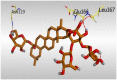 |
 |
| SAP8 |  |
 |
| N3, 13 |  |
 |