Fig. 3.
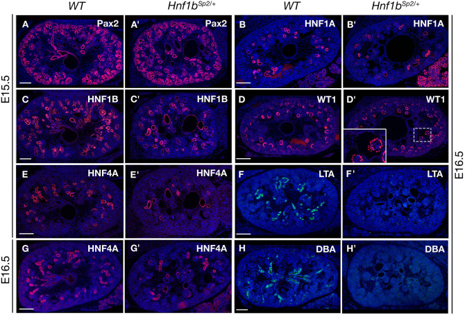
Hnf1bSp2/+ embryo kidneys exhibit normal ureteric bud branching but glomerular cysts and delayed proximal tubule (PT) differentiation. (A-H′) Immunohistochemical analysis of WT and Hnf1bSp2/+ embryo kidneys with Pax2 (A,A′), HNF1B (C,C′), the PT markers HNF4A (E,E′,G,G′), HNF1A (B,B′) and LTA (F,F′), WT1 (D,D′, inset shows glomerular cyst magnification with partially disorganized podocyte expression, and the collecting duct lectin DBA (H,H′) at the indicated stages. Note in Hnf1bSp2/+ sections decreased HNF4A+ PT structures (E′,G′) and PT dilatations, particularly at E15.5 (E′), which correlate with HNF1B expression in a serial section (C′) of the same embryo, decreased HNF1A PT expression together with increased acinar pancreatic expression (B′), and the absence of lectins LTA (F′) and DBA (H′). Sections are co-stained with 4′,6-diamidino-2-phenylindole (DAPI). Scale bars: 200 μm.

