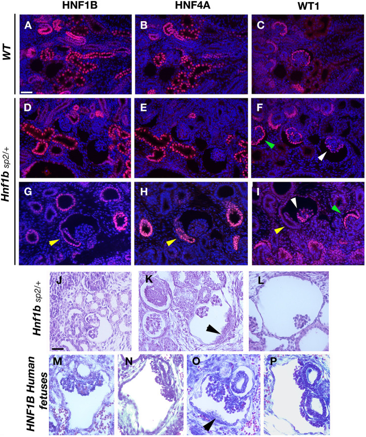
Fig. 4.
Glomerular cyst development, progressive tuft atrophy and abnormal PT implantation in Hnf1bSp2/+ embryos. (A-I) Histological comparison with human HNF1B mutant fetuses. Immunohistochemical analysis of E16.5 WT (A-C) and Hnf1bSp2/+ embryos (D-I). PTs are highlighted by the co-expression of HNF4A and HNF1B; developing glomeruli and condensed mesenchyme are shown by WT1 staining. Sections are co-stained with DAPI. Note that HNF4A staining in Hnf1bSp2/+ (E,H) was intentionally increased to better visualize positive PTs. Note in Hnf1bSp2/+ dilated PTs that were HNF4A+ (E,H), glomerular cysts with disorganized podocyte layer stained by WT1 (white arrowheads); non-cystic glomeruli show normal podocyte layer (green arrowheads). Yellow arrowheads (H,I) show abnormal lateral insertion of the glomerulotubular junction into Bowman's capsule and disorganized HNF4A expression (H). (J-P) H&E-staining of E17.5 Hnf1bSp2/+ embryos (J-L) and human mutant fetuses described in Haumaitre et al. (2006) (M-P). Black arrowheads indicate disorganized layer cells in mutants and human fetuses (K,O). Note also collapsed or disrupted tufts inside highly widened Bowman's capsules (L,P). Scale bars: 50 μm.

