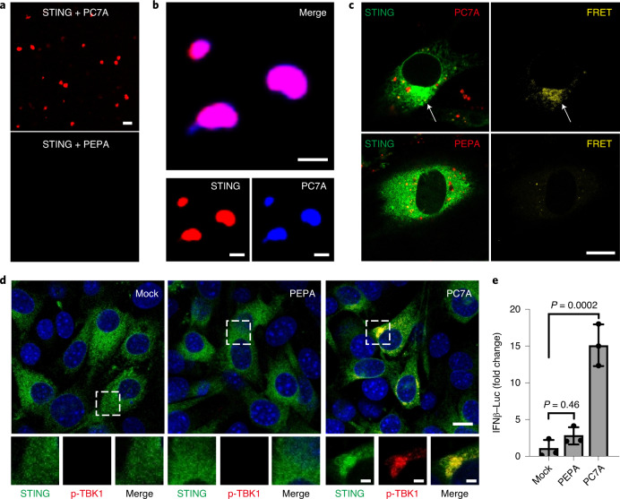
Fig. 2. PC7A polymer induces STING condensation and immune activation.
a, PC7A, but not PEPA, induces STING (Cy5-labelled) phase condensation after 4 h incubation. Scale bar, 10 μm. b, STING (4 μM, Cy5-labelled) and PC7A polymer (2 μM, aminomethylcoumarin acetate-labelled) are colocalized within the condensates. Scale bars, 5 μm. c, Hetero-FRET from GFP–STING to TMR–PC7A shows the colocalization of STING and PC7A in MEFs. Energy transfer was not observed from GFP–STING to TMR–PEPA. Cell culture conditions were identical to those described in Fig. 1. GFP (λex/λem = 488 nm/515 nm) and TMR (λex/λem = 555 nm/580 nm) signals are shown on the left in green and red, respectively. FRET signals (λex/λem = 488 nm/580 nm) are shown in yellow on the right. Scale bar, 10 μm. d, p-TBK1 is recruited into the STING–PC7A condensates. Scale bar, 10 μm. Insets: magnified images of the areas indicated by the white boxes. Scale bars, 2 μm. e, PC7A, but not PEPA, induces the expression of IFNβ–luciferase in ISG-THP1 cells. Data are mean ± s.d. n = 3 biologically independent experiments. Statistical analysis was performed using one-way analysis of variance (ANOVA). The confocal images in a–d are representative of at least three biologically independent experiments.

