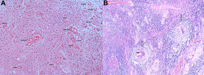
Figure 3.
Postoperative microscopy of the lesion showing (A) extensive coagulative necrosis of the mucosa with numerous broad, thin-walled, aseptate fungal hyphae surrounded by a prominent Splendore-Hoeppli phenomenon (marked with arrows) and (B) transmural dense chronic inflammatory infiltrates and multiple epithelioid cell granulomata with giant cells (marked with empty vertical arrows). Some giant cells are seen engulfing the fungal organism (marked with a solid horizontal arrow).

