Figure 4.
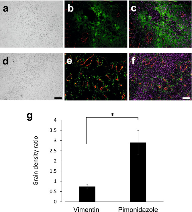
18F-FDG accumulation in hypoxic cells and host-derived cells. (a) Micro-autoradiography, (b) Merged image for pimonidazole (green) and CD31 (red), and (c) merged image for pimonidazole (green), CD31 (red), and grains (magenta) in the same view of the A431 cell-derived tumor section. (d) Micro-autoradiography, (e) Merged image for vimentin (green) and CD31 (red), and (f) Merged image for vimentin (green), CD31 (red), and grains (magenta) in the same view. (g) Grain density ratio of the pimonidazole-positive region to the CD31-positive region and the vimentin-positive region to the CD31-positive region (n = 3, mean ± SEM, Student’s t-test, two-sided, *: p < 0.05). Bars: 100 μm.

