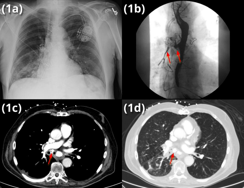
Figure 1.
(a) admission chest x-ray (CXR) indicating right lower lobe (RLL) pneumonia without evidence of broncholith on CXR
(b) modified barium swallow (MBS) demonstrating barium aspiration into the right mainstem bronchus (RMSB)
(c) mediastinal windows showing broncholith (arrow)
(d) lung windows showing both broncholith and post-obstructive RLL infiltrate

