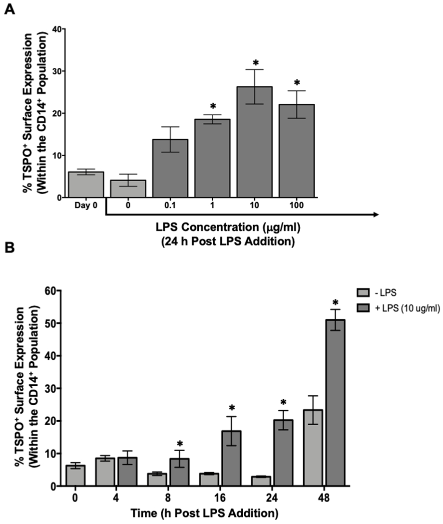
Figure 3. Human monocytes increase surface TSPO expression in a concentration- and time-dependent manner in response to LPS stimulation.

Human PBMC were isolated from leukocyte packs and stimulated with increasing concentrations of LPS (0.1–100μg/ml) for 24 hours, collected and stained for surface TSPO and CD14 expression. (A) Percentage of surface CD14+ and TSPO+ cells. (B) PBMC stimulated with 10μg/mL of LPS for different times. Average surface CD14 and TSPO at indicated time points of incubation. Results shown are the average of 2 independent experiments assessing a total of 6 human donors. * indicate significant differences at the p <0.05 level when compared to (A) Day 0 determined by a one-way ANOVA with a Dunnett’s multiple comparisons post-test and (B) No LPS at each time point as determined by an unpaired t-test. Values are mean ± SEM.
