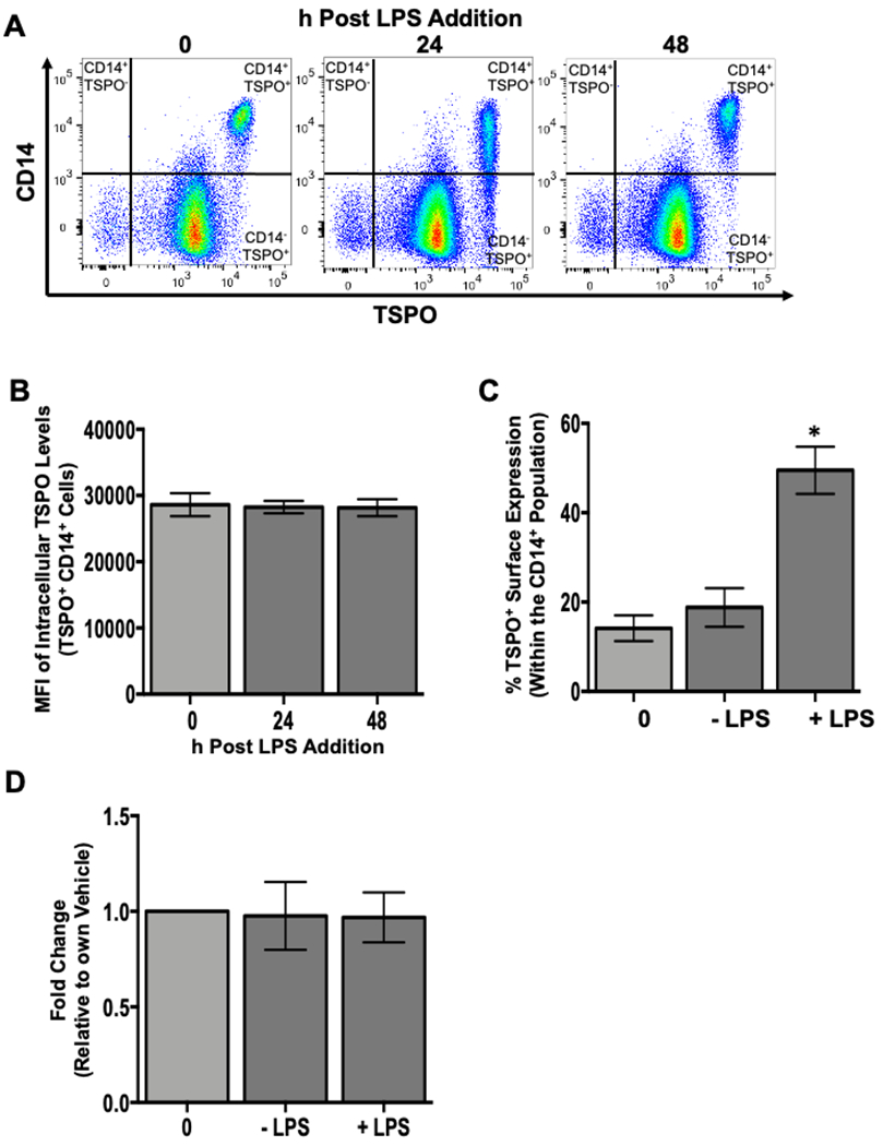
Figure 5. The increase in the percentage of monocytes with TSPO surface expression is not due to increased expression of intracellular TSPO or increased gene expression.

To assess whether the increased frequency of TSPO surface expression on monocytes following LPS treatment was due, in part, to increased intracellular expression, human PBMC were isolated and activated with LPS as described previously. At the indicated time points, cells were collected and stained for surface CD14, permeabilized, and stained intracellularly for TSPO. Purified monocytes were isolated and activated with LPS for 24 hours and stained for surface CD14 and TSPO to verify the increased frequency of TSPO surface expression following LPS treatment. Corresponding samples were also lysed at 24 hours post LPS treatment, mRNA was extracted from these isolated monocytes and qRT-PCR was used to determine TSPO gene expression. (A) Representative flow plots of intracellular TSPO staining at indicated time points. (B) Average mean fluoresence intensity (MFI) for intracellular TSPO. (C) Average increase in TSPO surface expression in purified monocytes. (D) Average fold change in TSPO mRNA expression in purified monocytes at 24 hours with and without LPS treatment. All data are from 2 independent experiments assessing a total of 6 human donors. * indicate significant differences at the p <0.05 level when compared to the time 0 control as determined by a one-way ANOVA with a Dunnett’s multiple comparisons post-test. Values are mean ± SEM.
