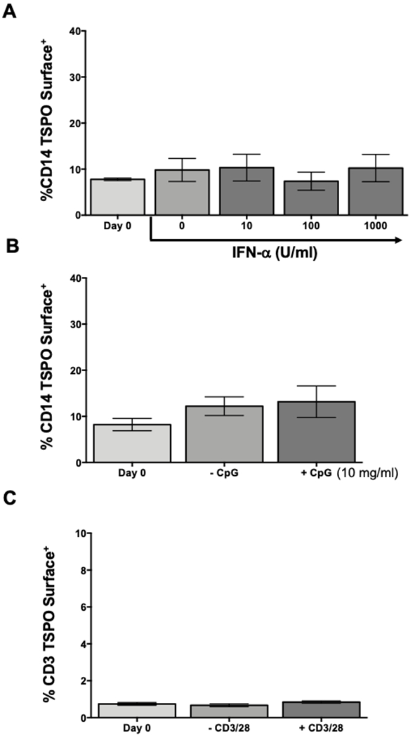
Figure 8. Increased percentage of TSPO surface expression on immune cells is not due solely to immune cell activation.

The data presented so far suggest that TSPO surface expression increases following monocyte activation with LPS. Next, we wanted to determine if this was a general hallmark of immune cell activation or if it was selective. To test this possibility, human PBMC were isolated as previously described and activated with either increasing concentrations of interferon α, 10μg/mL of CpG, or 5μg/mL of anti-CD3/CD28, a selective activator of T cells. Following 24 hours of activation, cells were collected and stained for surface CD14, CD3, or TSPO. (A) Frequency of TSPO surface expression on CD14 monocytes treated with increasing concentration of IFNα. (B) Frequency of monocyte TSPO surface expression after CpG activation. (C) Average increase of surface TSPO on CD3+ T cells after CD3/CD28 activation. Results are from 2 independent experiments assessing a total of 6 human donors. * indicate significant differences at the p <0.05 level compared to time 0 control as determined by a one-way ANOVA with a Dunnett’s multiple comparisons post-test. Values are mean ± SEM.
