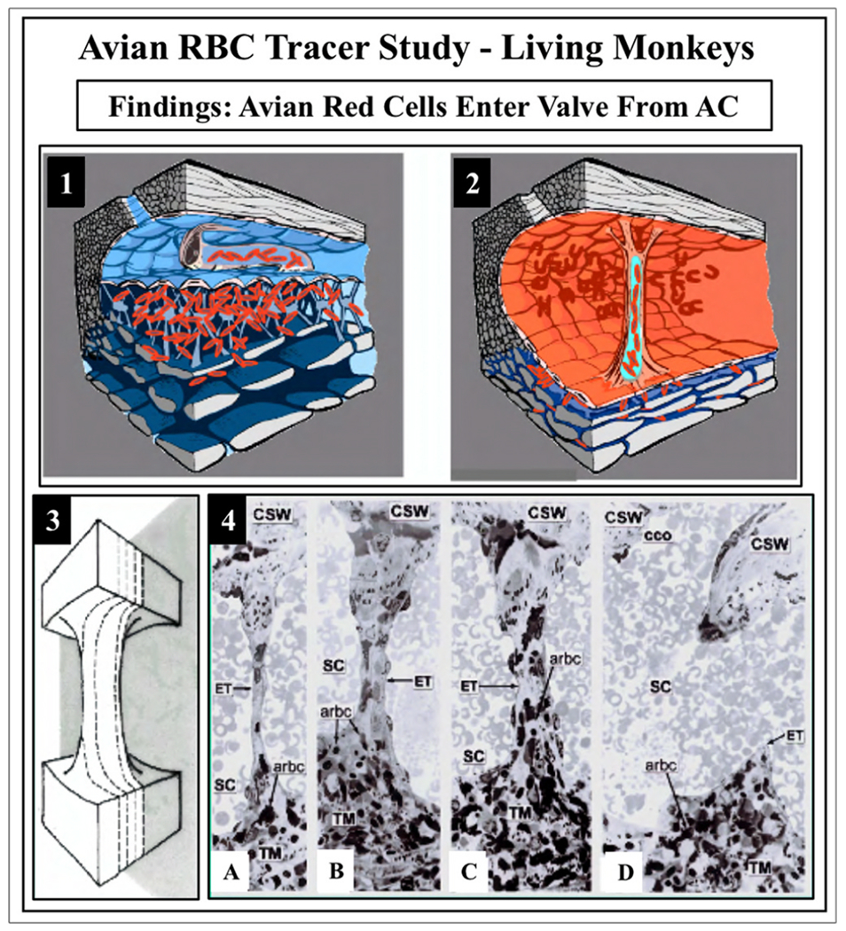
Fig. 14. Red blood cell tracers fill the lumen of Schlemm’s canal inlet valves.

Avian red blood cells (arbc) introduced into the anterior chamber (AC) as a tracer in living Macaca mulatto, monkey eye. (1) The arbc enter and fill a Schlemm’s canal (SC) inlet valve. (2) Gradual reduction of intraocular pressure (IOP) to 0 mm Hg in vivo causes SC to dilate and blood refluxes into the canal, because IOP is lower than episcleral venous pressure. Dilation of SC causes straightening and stretching of SC valves between SC walls enabling single radial sections to capture their full length. (3) Illustration of serial radial sections along a Schlemm’s canal inlet valve length. (4) From left to right: (A–D) are representative serial histologic sections encompassing the entire width of a Schlemm’s canal inlet valve depicted in (3). The endothelial lining (ET) of the SC valve is continuous with SC inner wall endothelium. The SC inlet valve spans across SC to attach to the corneoscleral wall (CSW). Avian red blood cells are present in the trabecular meshwork, and the juxtacanalicular space. In a central section through a Schlemm’s canal inlet valve, red cells fill the length of the lumen, as shown in C. In 4B and 4C, note the two collagenous supporting structures at the SC valve distal end with a narrow space where they meet. From Johnstone M, The aqueous outflow system as a mechanical pump, J Glaucoma 13, 421–438, 2004.
