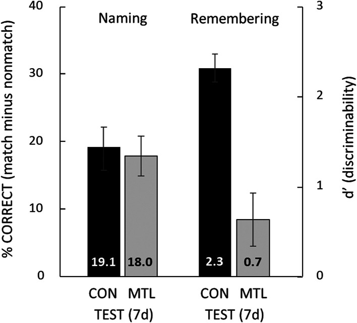
Fig. 6.
Remembering in contrast to naming. The percent correct naming scores are reproduced from the 7-d test in Fig. 4, which shows the amount of facilitation in naming (i.e., how much the naming of degraded images benefited from earlier presentation of their intact, matching images). G.P.’s score was 23.3%, the best of all the patients. For remembering (d′), the procedure at study and after 1 d was the same as for the naming test (Fig. 3). However, at 7 d after study, instead of taking another naming test, participants took a yes/no recognition memory test for the 40 old degraded images and 40 new degraded images. G.P. obtained a d′ score of 0.3 (55.0% correct), the poorest of all the patients. CON = 11 controls; MTL = 5 patients with MTL lesions. Error bars show SEM.

