Fig. 1.
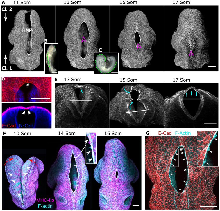
Surface ectoderm purse strings encircle the mammalian HNP. Representative whole-mount images of the developing cranial region in wild-type C57BL/6J mouse embryos. Throughout, zippering progression from Closure 2 is at the top, and progression from Closure 1 is at the bottom of the image. (A) Three-dimensional reconstructions of phalloidin-stained embryos illustrating the progression of HNP closure. Somite (Som) stages are as indicated. Magenta arrowheads indicate the direction of sectioning of the same embryos shown in E. (B and C) Stereoscope images of 11 Som (B) and 15 Som (C) stage embryo. The dashed green lines indicate regions where the NT is closed. (D, Top) Representative dorsal view of the C2z with the neuroepithelium stained with N-cadherin and surface ectoderm with E-cadherin. (Bottom) An optical reslice along the dashed line is shown below; arrowheads indicate the first point of contact, which is between surface ectoderm cells. (E) Transverse views into the same embryos indicated in A. White brackets indicate the distance between the lateral walls of the NT underlying the HNP. Cyan arrows indicate medial extension of a layer of cells, forming a thin closed layer by the 17 Som stage. (F) Dorsal views of representative embryos before (10 Som, * indicates the C1z) and after HNP formation. Actomyosin cable-like enrichments (arrows) are detected along the open neural folds. Red arrows indicate the necessary elevation of the neural folds, which precedes HNP formation. (Inset) Actin and myosin colocalization in the encircling cable (arrows). (G) Colocalization of cable F-actin with the surface ectoderm marker E-cadherin. (Inset) Membrane F-actin–rich ruffles (arrows) which appear to extend from the actomyosin cables.

