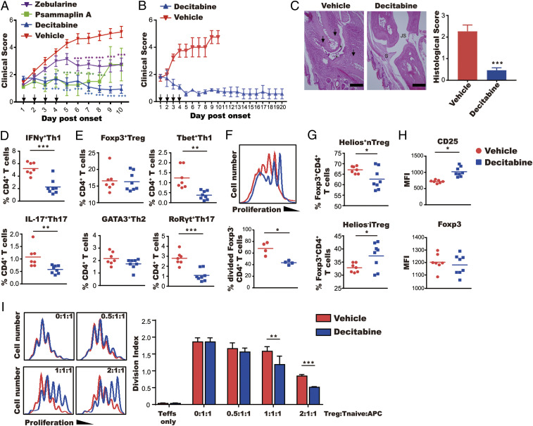
Fig. 1.
Decitabine has a sustained therapeutic effect on CIA. Mice with established arthritis were treated for 4 d with decitabine (1 mg⋅kg−1⋅d−1), psammaplin A (10 mg⋅kg−1⋅d−1), or zebularine (400 mg⋅kg−1⋅d−1). (A) Clinical scores (mean ± SEM; N = 10). (B) Clinical scores up to day 20 of mice treated with decitabine on days 1 to 4 (mean ± SEM; N = 7). (C) Representative images of proximal interphalangeal joint sections from mice treated with vehicle and decitabine are shown. Arrows indicate bone loss. Asterisks indicate damage of articular cartilage (JS, joint space; S, synovial membrane). (Scale bars, 100 μm.) The graph shows histological scores (mean ± SEM; N = 10). (D–H) Phenotypic characterization of Teff and Treg cells on day 10 of arthritis. Lymph node cells were stained with antibodies against cytokines (following stimulation with phorbol myristate acetate/ionomycin/brefeldin A) in D, lineage-specific transcription factors in E, or the nTreg cell marker Helios in G. Proliferation of FoxP3−CD4+ T cells was determined by CFSE labeling in F. Expression of FoxP3 and CD25 was quantified based on MFI in H. (I) Suppressive function of Treg cells was determined by culture with a fixed number of CFSE-labeled naive CD4+ T cells (CD25−CD4+) with mitomycin C-treated APCs from control mice under anti-CD3 antibody and IL-2 stimulation. Proliferation was determined by FACS (mean ± SEM; N = 3). *P < 0.05, **P < 0.01, ***P < 0.001.

