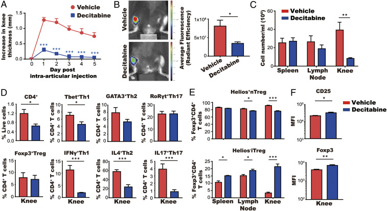
Fig. 5.
Accumulation of iTreg cells in the joint following treatment with decitabine. (A–C) Mice were immunized with mBSA and then treated for 5 d with decitabine (1 mg⋅kg−1⋅d−1) starting on day 10 after immunization (mean ± SEM; N = 5). Mice were given an intraarticular injection of mBSA 15 d after immunization. Knee swelling was monitored for 5 d (A). Cathepsin activity was measured using a fluorescent probe and detected using IVIS on day 2 of arthritis (B) and cell numbers in spleen, lymph node, and knee were counted on day 6 of arthritis (C). (D) Cells from arthritic joints were stained with antibodies against CD4+ T cell cytokines and lineage-specific transcription factors. Total numbers of cells are shown in SI Appendix, Fig. S4A. (E) Spleen, lymph node, and knee cells (following stimulation with mBSA) were stained with antibodies against the nTreg cell marker Helios. Total numbers of cells are shown in SI Appendix, Fig. S4B . (F) Expression of FoxP3 and CD25 of Treg cells gated from D was quantified based on MFI. *P < 0.05, **P < 0.01, ***P < 0.001.

