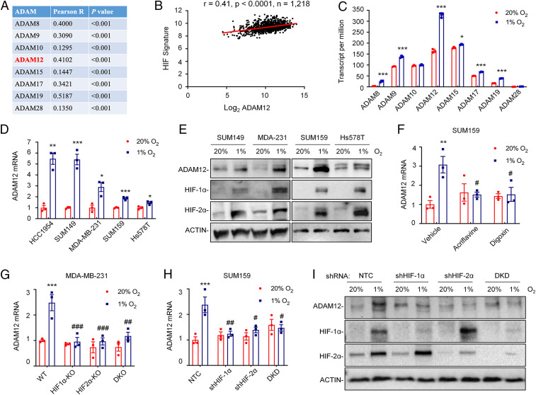
Fig. 1.
ADAM12 expression in breast cancer is correlated with HIF target gene expression and is induced by hypoxia in an HIF-dependent manner. (A and B) The mRNA expression of eight ADAM family members was compared with a 10-gene HIF signature in 1,218 human breast cancers from TCGA database by Pearson’s correlation test. For each comparison, the Pearson product-moment correlation coefficient (R = −1 to 1) and statistical significance (P) are shown (A). Individual data points for ADAM12 are shown (B). (C) RNA-seq data for the expression of eight ADAM family genes in SUM159 cells following exposure to 20% or 1% O2 for 24 h are shown (mean ± SEM; n = 3). *P < 0.05, ***P < 0.001 versus 20% O2 (Student’s t test). (D) RT-qPCR was performed to quantify ADAM12 mRNA levels in five human breast cancer cell lines following exposure to 20% or 1% O2 for 24 h. For each cell line, the expression of ADAM12 mRNA was quantified relative to 18S rRNA and then normalized to the result obtained from cells at 20% O2 (mean ± SEM; n = 3). *P < 0.05, **P < 0.01, ***P < 0.001 versus 20% O2 (Student’s t test). (E) Immunoblot assays were performed to determine ADAM12, HIF-1α, and HIF-2α protein levels in breast cancer cell lines following exposure to 20% or 1% O2 for 48 h. ACTIN was analyzed here (and elsewhere) as a loading control. (F) SUM159 cells were exposed to 20% or 1% O2 for 24 h in the presence of vehicle, digoxin (100 nM), or acriflavine (5 μM), and expression of ADAM12 mRNA was assayed by RT-qPCR. **P < 0.01 versus vehicle at 20% O2; #P < 0.05 versus vehicle at 1% O2 (two-way ANOVA with Tukey’s posttest). (G and H) RT-qPCR was performed to quantify ADAM12 mRNA levels in MDA-MB-231 (G) and SUM159 (H) subclones exposed to 20% or 1% O2 for 24 h. Data were normalized to WT (G) or NTC (H) cells at 20% O2 (mean ± SEM; n = 3). ***P < 0.001 versus WT or NTC at 20% O2; #P < 0.05, ##P < 0.01, ###P < 0.001 versus WT or NTC at 1% O2 (two-way ANOVA with Tukey’s posttest). (I) Immunoblot assays were performed to determine ADAM12, HIF-1α, and HIF-2α protein levels in whole cell lysates prepared from SUM159 subclones exposed to 20% or 1% O2 for 48 h. DKD, double knockdown; DKO, double knockout.

