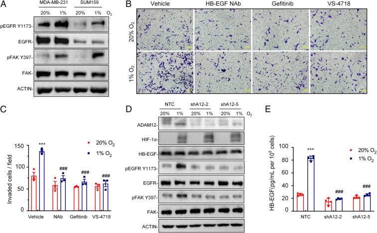
Fig. 6.
Hypoxia induces breast cancer cell invasion through ADAM12-EGFR-FAK signaling. (A) Immunoblot assays were performed to analyze phosphorylated EGFR (pEGFR Y1173), total EGFR, phosphorylated FAK (pFAK Y397), and total FAK protein in MDA-MB-231 and SUM159 subclone cells following exposure to 20% or 1% O2 for 24 h. (B and C) MDA-MB-231 cells were treated with vehicle, HB-EGF NAb (10 μg/mL), Gefitinib (5 μM), or VS-4718 (1 μM) and exposed to 20% or 1% O2 for 24 h. Cells that invaded through Matrigel-coated Boyden chamber inserts were stained with Crystal violet (B) and quantified by densitometry (C; mean ± SEM, n = 3). ***P < 0.001 versus cells exposed to vehicle at 20% O2; ###P < 0.001 versus cells exposed to vehicle at 1% O2 (two-way ANOVA with Tukey’s posttest). (D) MDA-MB-231 subclones stably transduced with NTC or ADAM12 shRNA vector (shA12-2 or shA12-5) were exposed to 20% or 1% O2 for 24 h, and whole-cell lysates were subjected to immunoblot assays. (E) MDA-MB-231 subclones stably transduced with NTC or ADAM12 shRNA vectors were exposed to 20% or 1% O2 for 72 h, and ELISA was performed to quantify HB-EGF in cell supernatant. Data shown are mean ± SEM, n = 3. ***P < 0.001 versus NTC cells at 20% O2; ###P < 0.001 versus NTC cells at 1% O2 (two-way ANOVA with Tukey’s posttest).

