
FIGURE 2.
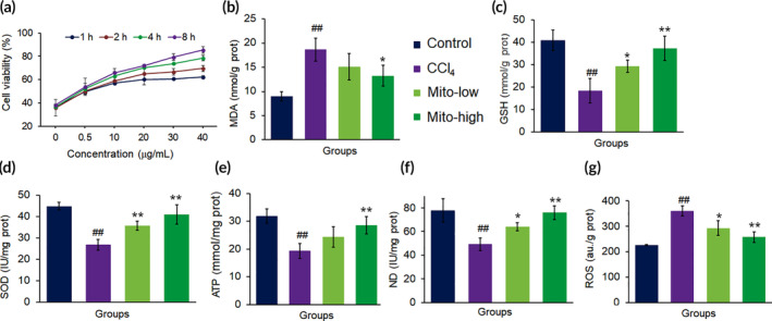
The mitochondria rescued hepatocytes damaged by CCl4. (a) Mitochondria increased cell viability in concentration‐ and time‐dependent manners. (b) MDA content decreased, whereas (c) GSH, (d) SOD, (e) ATP, and (f) ND increased in cell homogenates after the mitochondria were introduced into the cell media for incubation 8 h. Mito‐low, low concentration mitochondria (20 μg/ml); Mito‐high, high concentration mitochondria (40 μg/ml). Data were expressed as mean ± SD. ## p < 0.01 comparison with the normal control; *p < 0.05, **p < 0.01 compared with the CCl4 group. The values were averaged for six independent experiments