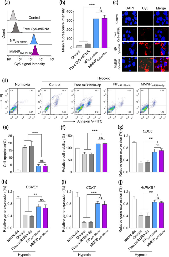
FIGURE 3.

NPmiR199a‐3p and MMNPmiR199a‐3p promote the proliferation of murine cardiovascular cells under hypoxic condition. (a) Flow cytometric analysis of HL‐1 cardiac muscle cell after 4 h incubation with free or nanoparticle encapsulated Cy5‐labeled miRNA. The concentration of Cy5‐miRNA was 100 nM. (b) Mean fluorescence intensity based on the flow cytometric analysis. Data represent means ± SD. ns, no significant difference. ***p < .001. (c) Confocal microscopic images show the cellular up‐taken of NPCy5‐miRNA and MMNPCy5‐miRNA. (d) Cell viability of HL‐1 cells after treated with different formulations. To generate hypoxic culture condition, HL‐1 cells culture was carried out in a hypoxic incubator containing 1% O2 for 6 h at 37°C. Cells were treated with free miR199a‐3p, NPmiR199a‐3p, and MMNPmiR199a‐3p (100 nM) for 24 h before hypoxic culture. Cell apoptosis and proliferation were examined after culture in hypoxic condition for 24 h. (d) and (e) Cell apoptosis were detected by flow cytometry after stained with PI and Annexin V‐FITC. (f) Cell viabilities were examined by MTT assay. The cell cycle related genes, including CDC6 (g), CCEN1 (h), CDK7 (i), and AURKB (j), were examined by real‐time PCR. Cells were treated as the MTT assay. Data represent means ± SD. ns, no significant difference. **p < .01, ***p < .001
