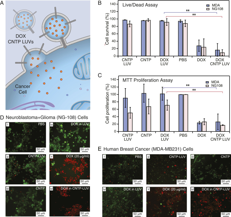
Fig. 3.
DOX delivery with CNTPs. (A) Schematic showing CNTP-LUV loaded with the DOX payload fusing to a cancer cell and delivering DOX to the cell interior. (B) Cell survival in live/dead assay after 48-h exposure of neuroblastoma-glioma (NG-108) and human breast cancer (MDA) cell cultures to DOX-CNTP-LUVs, CNTP-LUVs, free CNTPs, DOX-LUVs, free DOX, and PBS buffer (n = 9). **P 0.01. (C) Results of MTT cell proliferation assay after 48-h exposure of NG-108 and MDA cell cultures to DOX-CNTP-LUVs, CNTP-LUVs, free CNTPs, DOX-LUVs, free DOX, and PBS buffer. (NG-108 cells: n = 9; MDA cells: n = 15). **P 0.01. (D) Fluorescence microscopy images of NG108 cell culture with live and dead cells stained with green and red dye, respectively. Prior to imaging the cells were exposed for 48 h to (i) PBS buffer, (ii) CNTP-LUVs without the drug payload, (iii) CNTP solution, (iv) LUVs encapsulating DOX, (v) 20 g/mL of DOX, or (vi) CNTP-LUVs with encapsulated DOX. (E) Fluorescence microscopy images of MDA cell culture with live and dead cells stained with green and red dye, respectively. Prior to imaging the cells were exposed for 48 h to (i) PBS buffer, (ii) CNTP-LUVs without the drug payload, (iii) CNTP solution, (iv) LUVs encapsulating DOX, (v) 20 g/mL of DOX, or (vi) CNTP-LUVs with encapsulated DOX.

