Fig. 3.
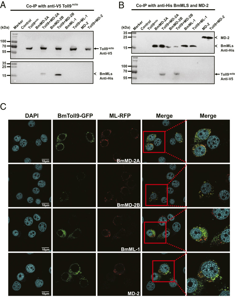
BmToll9 binds and colocalizes with BmMD-2A and BmMD-2B. (A and B) Outcomes of Co-IP experiments. Culture medium from untransfected BmN cells (control), or BmN cells expressing BmToll9ecto–V5 (Toll9ecto), BmMD-2A–His (BmMD-2A), BmMD-2B–His (BmMD-2B), BmML-1–His (BmML-1), or murine MD-2–His (MD-2) was collected alone or mixed. (A) Adding an anti-V5 antibody plus protein G beads to each sample immunoprecipitated BmToll9ecto from culture medium containing Toll9ecto alone or when medium containing Toll9ecto was mixed with each ML protein (Toll9 + BmMD-2A, Toll9 + BmMD-2B, Toll9 + ML-1, and Toll9 + MD-2) as detected by probing the immunoblot (Upper) with anti-V5. Probing the immunoblot (Lower) with anti-His shows that only BmMD-2A and BmMD-2B were coimmunoprecipitated. (B) Reciprocal experiment of adding anti-His antibody plus protein G beads to each of the samples. Anti-His plus protein G beads immunoprecipitated each of the ML proteins in the samples as detected by probing the immunoblot (Upper) with anti-His. Probing the immunoblot (Lower) with anti-V5 shows that only BmMD-2A and BmMD-2B coimmunoprecipitated with BmToll9ecto. (C) Confocal microscopy images showing BmN cells coexpressing recombinant BmToll9–GFP (green) plus BmMD-2A–RFP, BmMD-2B–RFP, BmML-1–RFP, or murine MD-2–RFP (red). BmToll9 colocalized with BmMD-2A and BmMD-2B on the surface of BmN cells as indicated by yellow in the merged images but did not colocalize with BmML-1 or MD-2. Nuclei were stained with DAPI. Cells were imaged 48 h posttransfection. (Scale bars, 10 μm.)

