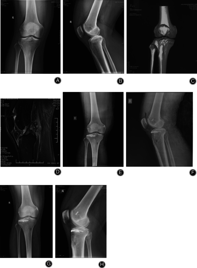
Fig 4.

Hyperextension bicondylar fracture. Male, 42 years old. (A, B) No fracture can be seen on X‐Ray. (C) 3D CT showed the compression of the anterolateral tibial plateau. (D) MRI showed the posterior cruciate ligament (PCL) injury. (E, F) The anterolateral articular surface was reduced and fixed with a horizontally‐oriented rim plate; arthroscopic reconstruction of the PCL was performed using the single‐bundle technique. (G, H) The fracture healed and satisfactory reduction had been achieved 1 year postoperatively.
