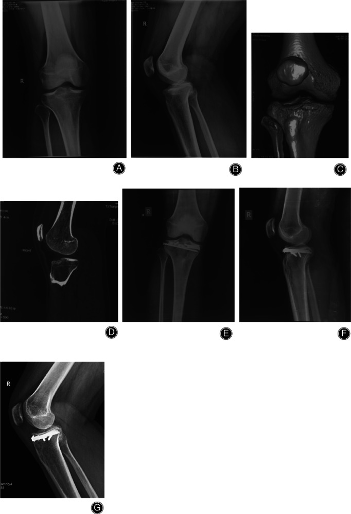
Fig 5.

Anteromedial + anterolateral tibial plateau fracture. Male, 33 years old. (A, B) X‐ray shows that no fracture could be seen. (C, D) 3D CT showed the compression of anteromedial + anterolateral tibial plateau, which was located behind the patellar tendon. (E, F) The fracture were reduced and fixed with a horizontally‐oriented rim plate. (G), The fracture healed and satisfactory reduction had been achieved 1 year postoperatively.
