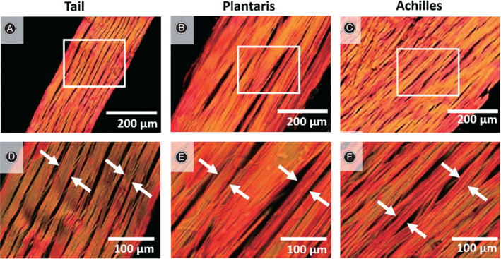
Fig 1.

Assessment of collagen fiber structure in longitudinal direction. (A‐C) Rat tail, plantaris, and Achilles tendons were cut in longitudinal direction, stained with picrosirius red, and showed at low magnification. White boxes represent areas observed at (D‐F) high magnifications, and collagen fibers are highlighted (white arrows). Adapted with permission from Lee et al. 33 .
