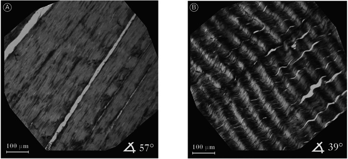
Fig 2.

Typical spatial distribution of local orientations of collagen fibers in Achilles tendons, resolution 1 × 1 pixel. Image (A) shows the area where nearly all collagen fiber bundles are aligned at 57°. In contrast, image (B) depicts a specimen of Achilles tendon containing undulated collagen fibers. Adapted with permission from Novak et al. 43 .
