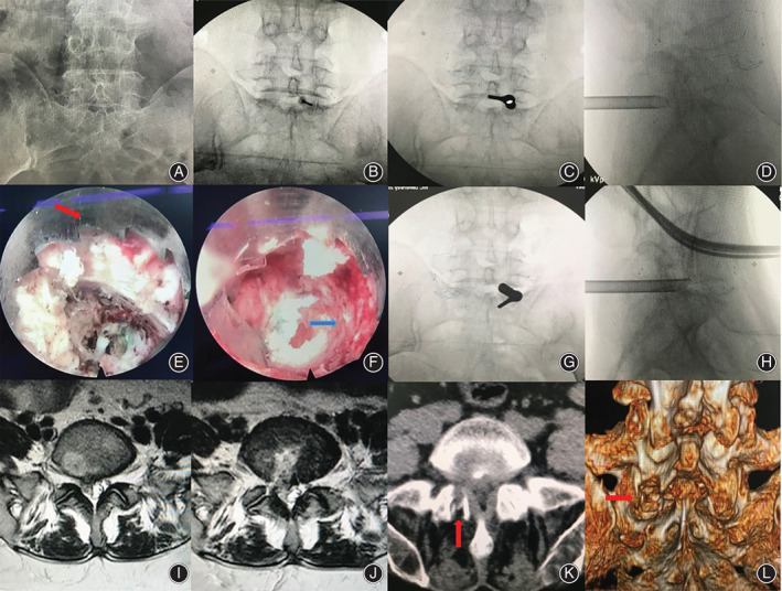
Fig. 6.

Typical case three. (A) Lumbar anterior–posterior X‐ray image. Narrow interlaminar window of L5–S1 level. (B, C, and D) The target puncture point and working cannula were located at the safety zone. (E and F) Endoscopic visible trephine (red arrow) was used for the laminoplasty (blue arrow). (G and H) Working cannula reached herniated disc tissue via the spinal canal under endoscopic observation. (I and J) Preoperative and postoperative MRI showed removal and good decompression of the S1 nerve root and dura. (K and L) Postoperative CT scan revealed laminoplasty (arrow).
