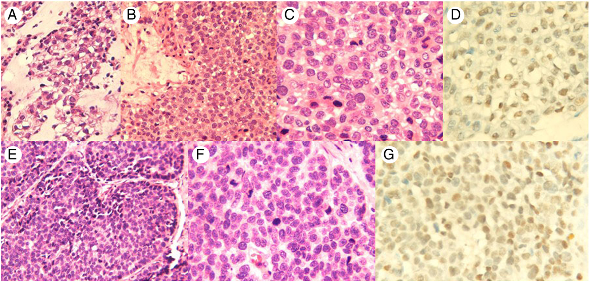
Fig. 1.

Case 1, A-D, The tumor cells show focal clear cell change and chondromyxoid background (H&E, A and B 200×; C, 400×); SOX10-positive triple-negative breast cancer (D, SOX10 IHC, 400x,). Case 2, E-G, The tumor cells are poorly differentiated and are arranged in solid nests (H&E, E, 200×; F, 400×); SOX10-positive triple-negative breast cancer (G, SOX10 IHC, 400×).
