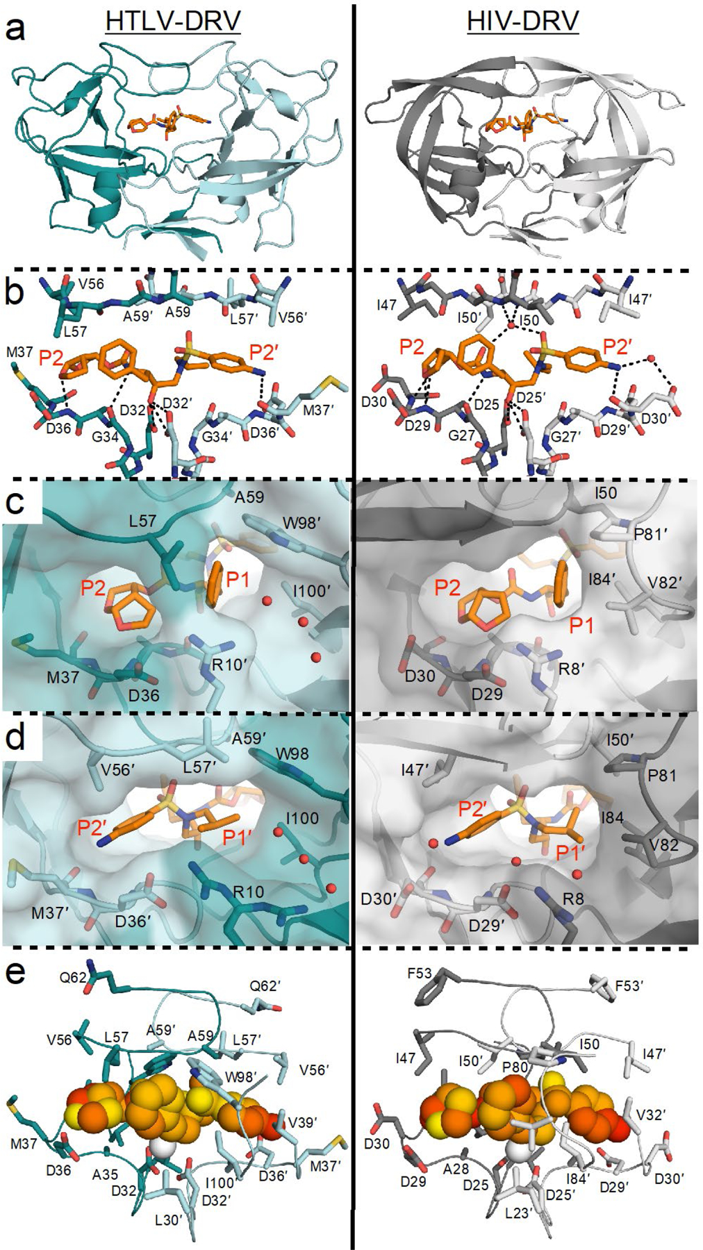
Figure 2. Comparison of DRV binding to HTLV-1 versus HIV-1 protease.

(a) Cocrystal structures of DRV bound to HTLV-1 and HIV-1 protease (PDB IDs 6W6Q and 6DGX, respectively). (b) Inter-molecular hydrogen bonds between DRV and protease active site. (c) Close-up view of aniline moiety in the S2′ subsite. (d) Close-up view of bis-THF moiety in the S2 subsite. (e) Packing around DRV at the active site visualized through mean inter-molecular vdW interactions with protease during MD simulations, where yellow to red indicates increased packing. In all panels, the prime side monomer (chain A) interacting with the bis-THF moiety is in darker shade, and DRV is depicted as orange sticks in panels A–D.
