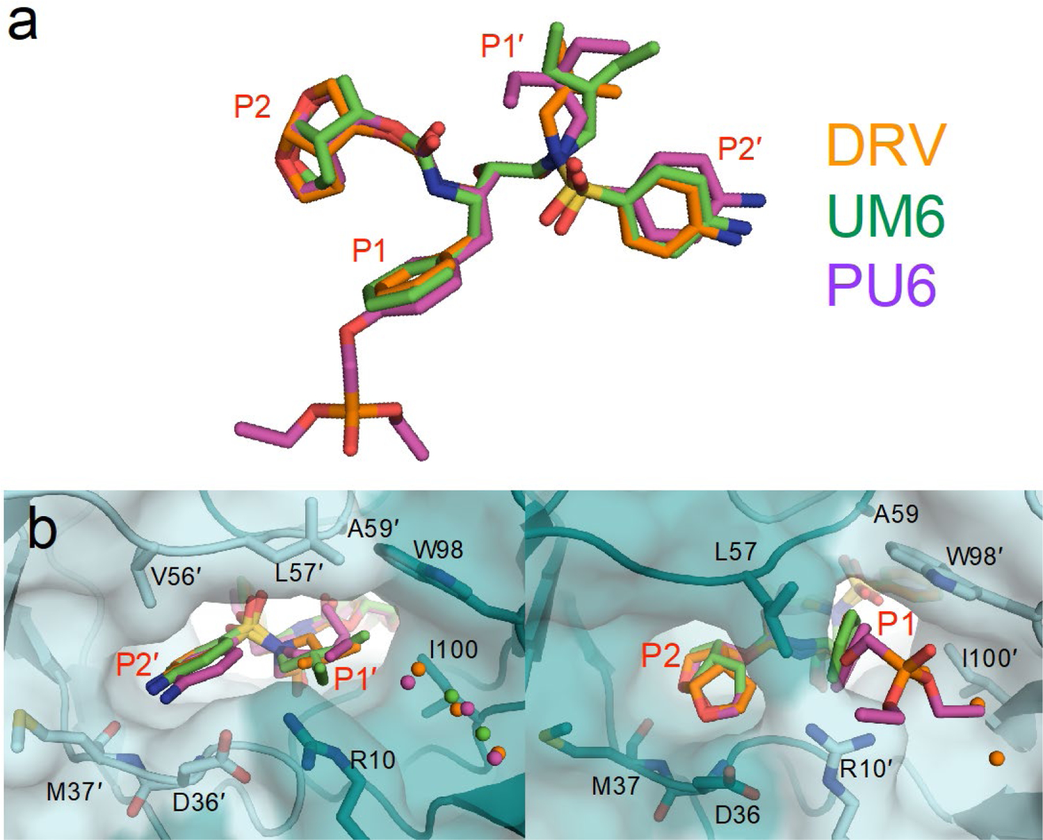
Figure 3.

Comparison of DRV and DRV analogs when bound to HTLV-1 protease. (a) Alignment of inhibitors. (b) Close-up view of P2′–P1′ moiety in the S2′–S1′ subsite, and P2–P1 moieties in the S2–S1 subsite. The phosphonate moiety of PU6 extends into the S1 subsite, displacing conserved crystallographic waters.
