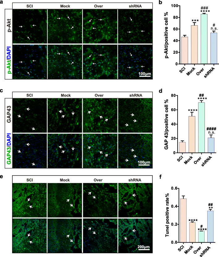
Fig. 6.
CD157 overexpression in BMSCs attenuates inflammation and apoptosis of the injured neurons in vivo (2). a, c Representative immunofluorescence images of p-Akt and GAP 43 of neurons at the injured site. The white arrows indicate the positive cells. Scale bars, 100 μm. b, d Statistics of Grp 78 and GAP 43 positive rate. n.s., non-significant, ****p < 0.0001 vs SCI group. #p < 0.05, ##p < 0.01, ###p < 0.001, ####p < 0.0001 vs Mock group. e TUNEL staining images of neurons at the damage site. The white arrows indicate the positive cells. Scale bars, 200 μm. f Statistics of TUNEL positive rate. **p < 0.01, ****p < 0.0001 vs SCI group. #p < 0.05, ##p < 0.01 vs Mock group

