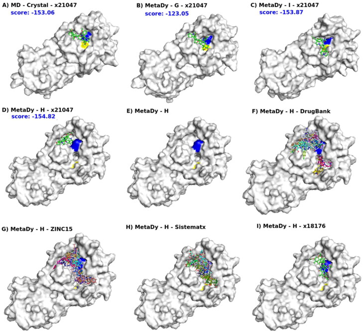
Figure 12.
Multi-conformer flexible docking’s hits explore the MCov2Pro active site space in different ways. For all, the blue and yellow patches on the molecular surface indicate the presence of the catalytic dyad (His41 and Cys145, respectively). In (A), a representative conformer of MD at equilibrium from the crystallographic structure (MD - crystal). In (B, C), the representative conformers G and I from a well-tempered MetaDy, respectively. In (D), the representative conformer H from a nontempered MetaDy. Conformers G, I and H are in increasing order of RMSD in relation to the crystallographic structure (see Figure 10). Also, from (A) to (D) we show the docking of the same ligand x21181 into MD crystal, G, I and H conformes, with respective scores in blue. In (E), the H conformer highlighting its bicavitary site. Note that the dyad residues are separated and this opens a cavity in between. From (F) to (H), superimposed docking of several ligands selected from DrugBank, ZINC15 and Sistemamatx, respectively. In (I), an example of ligand (x18176) that binds both cavities of the H conformer. (See Figure 10).

