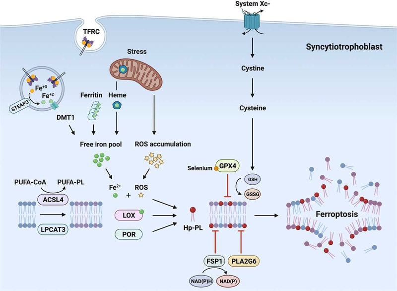
Figure 1. A schematic depicting key pathways in ferroptosis, delineated in this review.
The text cites direct evidence for the accumulation of Hp-PE, a main form of pro-ferroptosis Hp-PL, in placental injury. The role of GPX4 and PLA2G6 in mitigating trophoblast ferroptosis is also highlighted. The figure also depicts key sources of trophoblastic iron pool. Additional, indirect evidence supports the presence of most ferroptotic regulators in human trophoblasts. Abbreviations: ACSL4, acyl-CoA synthetase long-chain family member 4; DMT1, divalent metal transporter 1; FSP1, ferroptosis suppressor protein 1; GPX4, glutathione peroxidase 4; GSH, glutathione; GSSG, oxidized glutathione; Hp-PE, hydroxy-peroxidized phosphatidylethanolamine; LOX, lipoxygenase; LPCAT3, lysophosphatidylcholine acyltransferase 3; NAD(P), Nicotinamide adenine dinucleotide phosphate; PLA2G6, phospholipase A2 Group VI; PUFA, polyunsaturated fatty acid; PUFA-PE, phosphatidylethanolamine-containing polyunsaturated fatty acid chain; POR, cytochrome P450 oxidoreductase; ROS, reactive oxygen species; STEAP3, six-transmembrane epithelial antigen of prostate 3; TFRC, transferrin receptor

