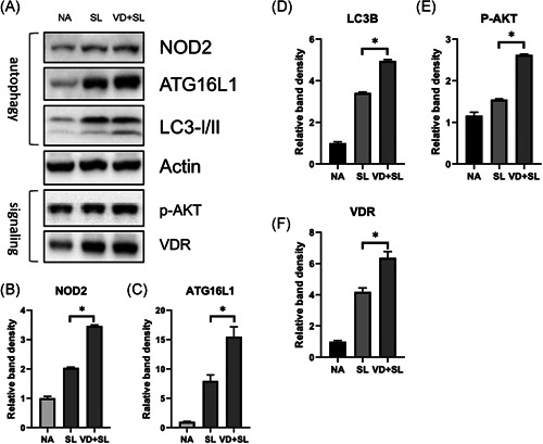
Figure 4.

Effect of 1,25D3 treatment on the expression of cecal proteins in Salmonella colitis mice. Female 6–8‐week‐old C57BL/6 mice were administered with streptomycin, and incubated with PBS (NA) or 108CFU of ST (SL1344 strain, SL) for 48 h, along with 1,25D3 (VD + SL) daily for 14 days. The cecum homogenates were used to assess the protein expression by immunoblotting with antibody to detect NOD2, Atg16L1, LC‐3B, VDR, and activated AKT (p‐AKT) proteins; actin was used for normalization of total protein. Representative immunoblots (A) and densitometric quantification of immunoreactive bands (B−F) are shown. The relative band intensities of NOD2 (B), Atg16L1 (C), LC‐3B (D), p‐AKT (E), and VDR (F) in protein extracts of cecal tissues in Salmonella colitis mice in the presence or absence of 1,25D3 were quantified as fold increases compared with those of the control mice. Data are expressed as mean ± SEM (n = 3 mice per group). *p < .05. 1,25D3, 1,25‐dihydroxyvitamin D3; Atg16L1, autophagy‐related 16 like 1; NOD2, nucleotide‐binding oligomerization domain‐containing protein 2; PBS, phosphate‐buffered saline; ST, Salmonella enterica serovar Typhimurium; VDR, vitamin D receptor
