
Figure 1.
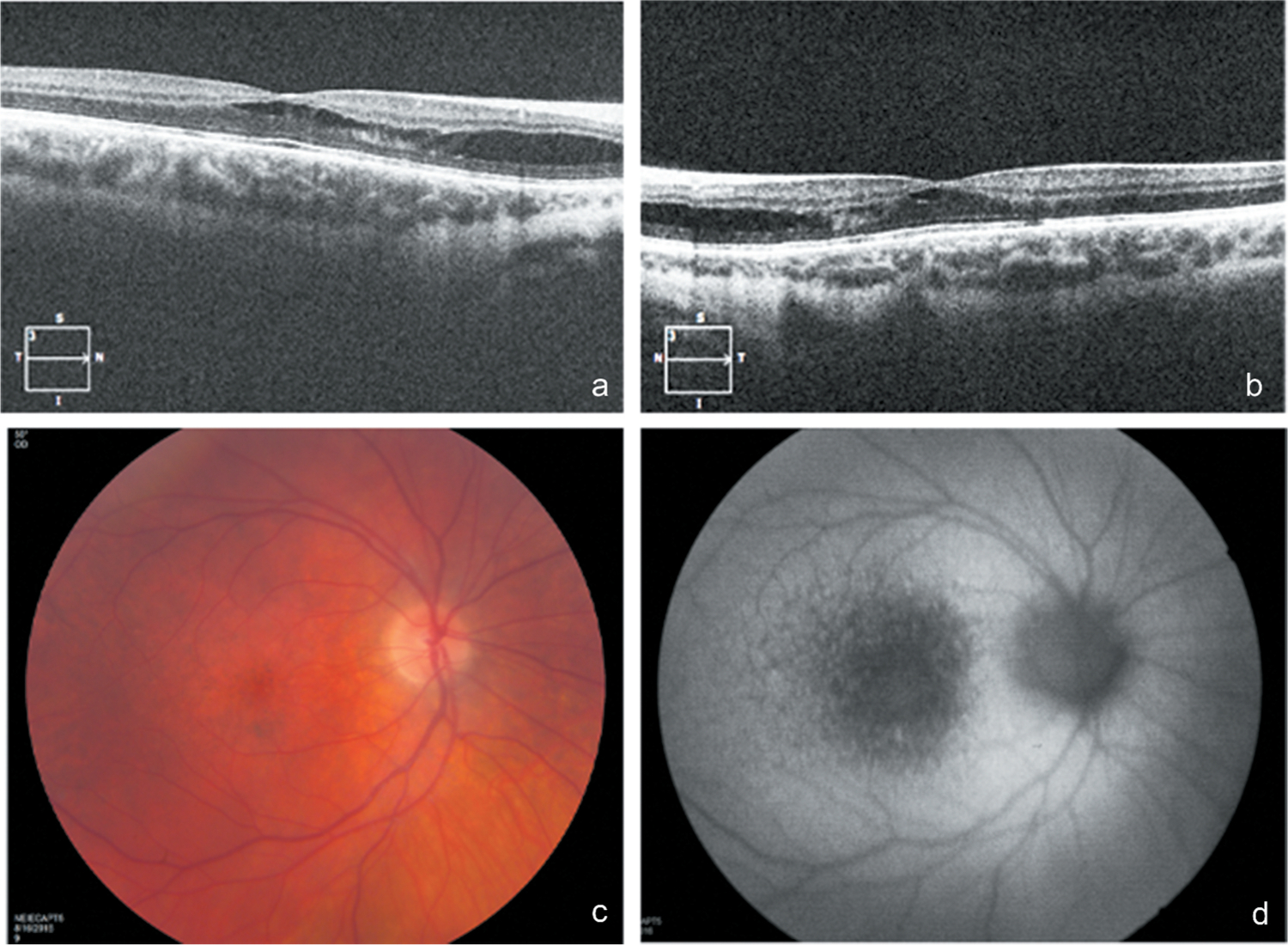
Spectral-domain OCT image of the retina through the fovea of the right (a) and left (b) eyes demonstrating retinoschisis. To highlight details of the schisis cavity, the contrast was increased from the original images. Fundoscopy demonstrated mild pigment mottling of the macula (c), better appreciated as hypoautofluorecent stippling on blue light autofluorescence (d).