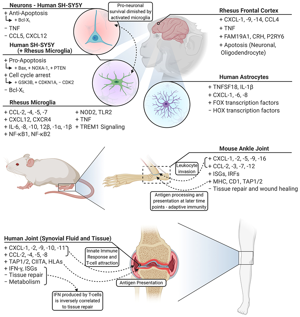
Fig. 4.

An overview of the transcriptional response of late disseminated infection. Following hematogenous dissemination, the central nervous system and joints are common regions for B. burgdorferi s.l. to colonize. Frontal cortex tissue responds in a diverse manner representative of resident cells – highlighted by inflammatory cytokines and marked by neuronal and oligodendrocyte apoptosis. SH-SY5Y neuronal cells indicate minimal response to infection and are inclined towards a pro-survival profile. Through the activation of microglia, marked by inflammatory factors, SH-SY5Y cells shift towards cell cycle arrest and pro-apoptotic profiles. Astrocytes promote a less robust immune response compared to microglia and show a shift in transcriptional regulation. B. burgdorferi invade the synovial fluid and surrounding tissue of joints, leading to the induction of chemotactic cytokines and subsequent invasion of leukocytes – an indication of Lyme arthritis. Created with BioRender.com.
